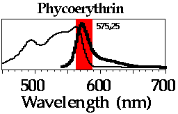

The excitation spectrum and emission spectra (bold line) are shown for
PE. The red-shaded region represents the bandpass filter commonly used to
measure PE fluorescence. On most flow cytometers, PE is excited by an argon
laser tuned to 488 nm; however, much more sensitive detection is obtained
with a laser tuned to 533 nm.
Also see the absorbance spectrum for PE.
Compare the PE fluorescence spectra to spectra for
several FACS immunofluorescence dyes.